

Laser Scanning Microscopes
SKU: ME-LE-46
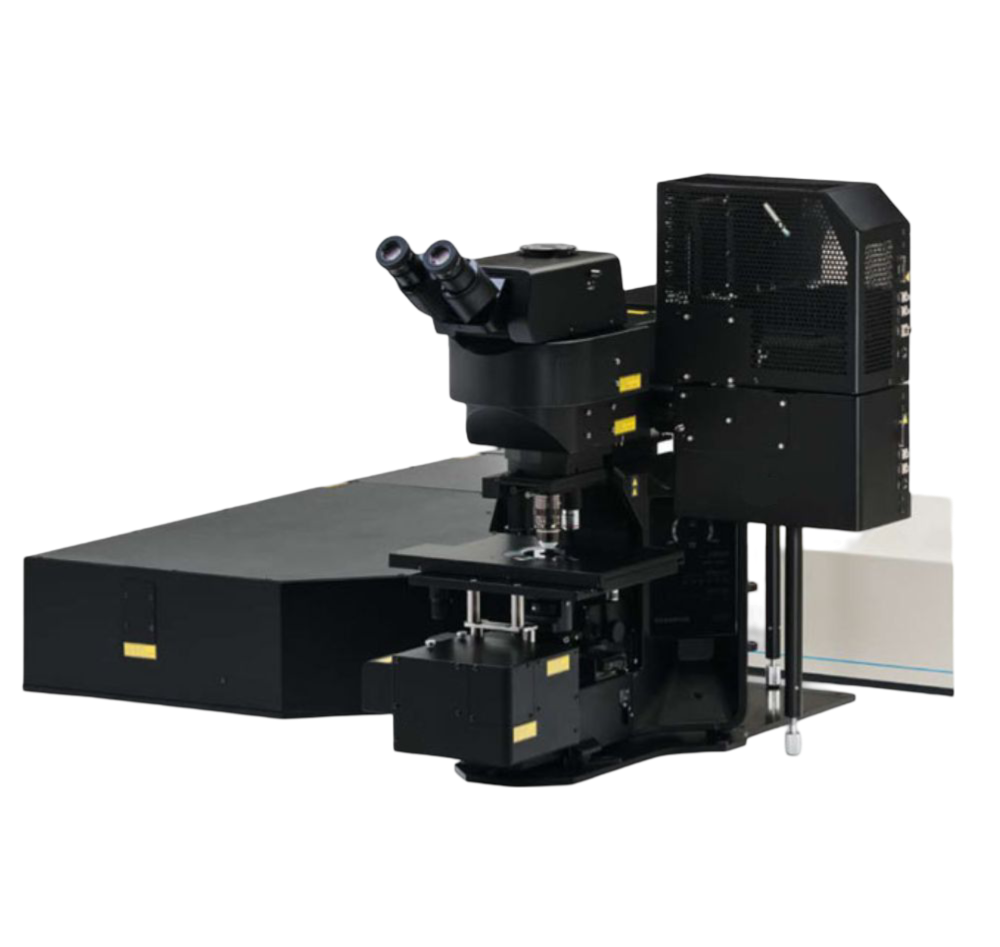
If you're in search of precision and clarity in your medical imaging, look no further than our Laser Scanning Microscopes. These cutting-edge microscopes utilize laser beams to provide incredibly high-resolution images of biological samples, making them an invaluable tool for medical research and diagnosis. With advanced 3D imaging capabilities, these microscopes allow for the detailed visualization of cells, tissues, and processes within the body. Our laser scanning microscopes are designed to be user-friendly, with intuitive software and easy-to-use controls, making them perfect for medical professionals of all experience levels. Whether you're studying cellular structures or examining diseased tissues, our laser scanning microscopes will provide you with the accurate and detailed images you need for your medical work.
- Biomedical Research: LSMs are extensively used in biomedical research for studying various biological specimens such as cells, tissues, and even whole organisms. They provide detailed three-dimensional images of cellular structures, helping researchers understand cellular processes, organelle dynamics, and interactions between molecules.
- Neuroscience: LSMs play a crucial role in neuroscience research by enabling high-resolution imaging of neuronal structures and neural circuits. They are used to visualize dendritic spines, axonal processes, synaptic connections, and neuronal activity in live brain tissue, providing valuable insights into brain function and development.
- Materials Science: In materials science, LSMs are used to investigate the microstructure and surface morphology of materials at the nanoscale. Researchers use LSMs to study defects, grain boundaries, and interfaces in materials such as metals, ceramics, polymers, and composites, facilitating the development of new materials with improved properties.
- Confocal Microscopy: LSMs are widely employed in confocal microscopy, a technique that enhances image contrast and resolution by eliminating out-of-focus light. Confocal LSMs are used in various fields, including biology, medicine, and materials science, for imaging fluorescently labeled specimens with high spatial resolution and optical sectioning capabilities.
- Live Cell Imaging: LSMs equipped with live cell imaging capabilities are used to study dynamic cellular processes in real time. By capturing time-lapse images of living cells, researchers can observe cellular events such as cell division, migration, and signaling pathways, shedding light on fundamental biological processes and disease mechanisms.
- Drug Discovery and Development: LSMs are utilized in drug discovery and development for screening potential drug candidates and studying their effects on cellular structures and functions. High-content screening systems based on LSMs enable researchers to analyze multiple cellular parameters simultaneously, accelerating the identification of novel therapeutic compounds.
- Biophysics and Bioengineering: LSMs are employed in biophysics and bioengineering studies to investigate the mechanical properties and biomechanics of biological tissues and cells. By combining LSMs with techniques such as atomic force microscopy (AFM) or optical tweezers, researchers can measure forces exerted by cells, study cell-substrate interactions, and explore tissue mechanics at the microscale.
- Environmental Science: LSMs are used in environmental science for studying microorganisms, pollutants, and nanoparticles in environmental samples. They help researchers visualize microbial communities, track pollutant degradation processes, and investigate interactions between nanoparticles and environmental matrices, contributing to our understanding of environmental processes and pollution remediation strategies.